
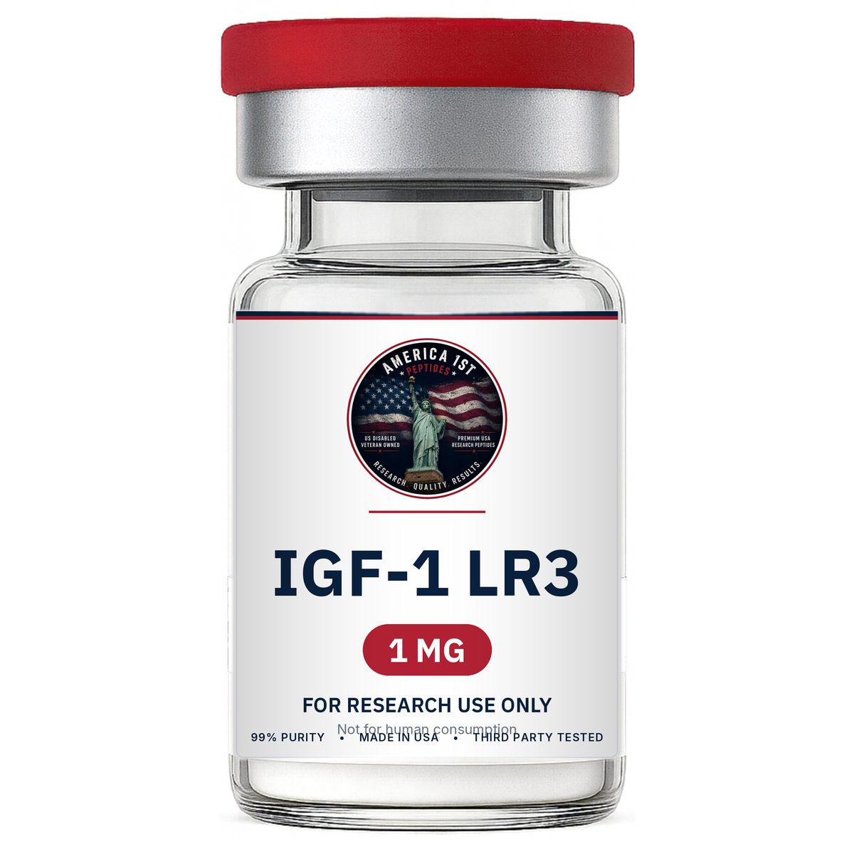
Description
What is IGF1-LR3?
IGF1-LR3 (Insulin-like Growth Factor-1 Long Arg3) is a synthetic, extended version of naturally occurring IGF-1. This modified peptide differs from native IGF-1 by the addition of 13 amino acids at the N-terminal and the substitution of glutamic acid at position 3 with arginine. These structural modifications dramatically reduce its affinity for IGF-binding proteins, allowing IGF1-LR3 to remain active in the bloodstream up to 120 times longer than standard IGF-1.
As a result, IGF1-LR3 plays a vital role in stimulating cell proliferation, differentiation, repair, and communication, with significant effects in muscle, bone, nerve, liver, kidney, and connective tissues.
IGF1-LR3 Structure

-
Sequence: MFPAMPLSSL FVNGPRTLCG AELVDALQFV CGDRGFYFNK PTGYGSSSRR APQTGIVDEC CFRSCDLRRL EMYCAPLKPA KSA
-
Molecular Formula: C400H625N111O115S9
-
Molecular Weight: 9117.5 g/mol
-
CAS Number: 946870-92-4
-
Source: PubChem
Research Areas
Cell Division and Proliferation
IGF1-LR3 is a potent stimulator of hyperplasia (new cell formation) rather than hypertrophy (cell enlargement). It drives connective tissue growth, including muscle and bone, while also supporting regeneration in liver, kidney, nerve, skin, and vascular tissues. Because of its prolonged half-life, IGF1-LR3 provides up to 3x the activation of native IGF-1 per dose, making it a superior research molecule for tissue development studies.

Metabolic Function and Diabetes Research
IGF1-LR3 interacts with both IGF-1 receptors and insulin receptors, increasing glucose uptake in muscle, nerve, and liver cells. This leads to lower blood sugar, enhanced glycogen breakdown, and fat utilization. Animal studies suggest IGF1-LR3 may reduce insulin requirements by approximately 10%, highlighting potential applications in diabetes and metabolic research.
Myostatin Inhibition and Muscle Preservation
Myostatin naturally limits muscle cell growth and repair. IGF1-LR3 counteracts this effect by activating MyoD, a protein central to muscle regeneration and hypertrophy. In animal models of Duchenne Muscular Dystrophy (DMD), IGF1-LR3 demonstrated protective effects against muscle cell apoptosis and breakdown, making it a candidate for studies on muscle-wasting conditions.
Longevity and Anti-Aging Research
IGF1-LR3 has been linked to tissue repair, mitochondrial protection, and cellular maintenance. Research in mammals suggests it may play a role in delaying degenerative processes and extending lifespan by reducing disability and promoting regenerative signaling. Ongoing studies in mice explore its potential against dementia, kidney disease, and muscle atrophy.
Glucocorticoid Side-Effect Mitigation
Long-term glucocorticoid use often leads to muscle wasting, fat accumulation, and bone density loss. Preliminary studies indicate IGF1-LR3 may help offset these side effects, offering a protective role in long-term steroid therapies.
Disclaimer
IGF1-LR3 exhibits minimal to moderate side effects in animal models and demonstrates low oral but excellent subcutaneous bioavailability. Dosages observed in animal research do not scale to humans.
All IGF1-LR3 offered by America 1st Peptides is provided strictly for educational and scientific research use only. These products are not medicines or drugs, have not been evaluated or approved by the FDA, and are not intended for human or veterinary use.
Referenced Citations
-
“Adipose Tissue-Derived Stem Cell Secreted IGF-1 Protects Myoblasts from the Negative Effect of Myostatin.” Hindawi, 2014.
-
Li, N. et al. “Myostatin Attenuation In Vivo Reduces Adiposity, but Activates Adipogenesis.” Endocrinology, 2016.
-
Corpas, E. et al. “Human Growth Hormone and Human Aging.” Endocr. Rev., 1993.
-
Sonntag, W. et al. “Diverse Roles of Growth Hormone and IGF-1 in Mammalian Aging.” J. Gerontol., 2012.
-
NCBI. “IGF-I/IGFBP system: metabolism outline and physical exercise.”
-
Hanaoka, B.Y. et al. “Implications of Glucocorticoid Therapy in Idiopathic Inflammatory Myopathies.” Nat. Rev. Rheumatol., 2012.
-
Philippou, A. et al. “The Role of IGF-1 in Skeletal Muscle Physiology.” J Musculoskelet Neuronal Interact, 2007.
-
Philippou, A. et al. “Expression of IGF-1 Isoforms after Exercise-Induced Muscle Damage.” In Vivo, 2009.
Reviews
There are no reviews yet.